Admin
مدير المنتدى


عدد المساهمات : 18726
التقييم : 34712
تاريخ التسجيل : 01/07/2009
الدولة : مصر
العمل : مدير منتدى هندسة الإنتاج والتصميم الميكانيكى
 |  موضوع: كتاب CT of the Airways موضوع: كتاب CT of the Airways  السبت 02 أبريل 2022, 5:56 pm السبت 02 أبريل 2022, 5:56 pm | |
| 
أخواني في الله
أحضرت لكم كتاب
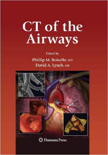
CT of the Airways
Edited by
Phillip M. Boiselle, MD
Beth Israel Deaconess Medical Center and Harvard Medical School, Boston, MA
and
David A. Lynch, MB
National Jewish Medical and Research Center, Denver, CO

و المحتوى كما يلي :
Contents
Series Editor’s Introduction . v
Preface ix
Contributors xiii
Part I Introductory
1 Airway Anatomy and Physiology 3
Kathryn Chmura, Stella Hines, and Edward D. Chan
2 Radiologic Anatomy of the Airways 25
Phillip M. Boiselle and David A. Lynch
3 Pathology of the Airways . 49
Chih-Wei Wang and Thomas V. Colby
4 Anatomical Airway Imaging Methods 75
Phillip M. Boiselle and David A. Lynch
5 Functional Airway Imaging Methods . 95
Jonathan G. Goldin
Part II Large Airways
6 Tracheobronchial Stenoses 121
Phillip M. Boiselle, Jay Catena, Armin Ernst, and David A. Lynch
7 Tracheal and Bronchial Neoplasms . 151
Karen S. Lee and Phillip M. Boiselle
8 Tracheobronchomalacia . 191
Ronaldo Hueb Baroni, Rodrigo Caruso Chate, Daniel Nobrega da Costa,
and Phillip M. Boiselle
9 Bronchiectasis 213
John D. Newell, Jr.
Part III Small Airways
10 Asthma . 239
Philippe A. Grenier, Catherine Beigelman-Aubry, and Pierre-Yves Brillet
11 Infectious Small Airways Diseases and Aspiration Bronchiolitis 255
Kyung Soo Lee
12 Noninfectious Inflammatory Small Airways Diseases . 271
David A. Lynch
13 Obliterative Bronchiolitis . 293
C. Isabela S. Silva and Nestor L. Müller
14 Smoking-Related Small Airways and Interstitial Lung Disease 325
David M. Hansell and Athol U. Wells
xixii Contents
Part IV: Pediatric Airways Disorders
15 Large Airways 351
Edward Y. Lee and Marilyn J. Siegel
16 Computed Tomography of Pediatric Small Airways Disease 381
Alan S. Brody
Index .
Index
Acute respiratory distress syndrome (ARDS), 314
Adenoid/nasopharyngeal tonsil, 5
Adenoid cystic carcinoma, 155, 157, 159,
171–175
Air trapping
and expiratory CT, 46, 89, 111, 243–245, 300
Airway malignant lesions
adenoid cystic carcinoma, 173–174
carcinoid tumors, 175–177
mucoepidermoid carcinomas, 177–179
squamous cell carcinoma, 168–171
Airways
conducting portion of and terminal bronchioles, 12
histology of, 11
innervation, 10–11
lipomas, 164–165
measurement of, 14–16, 32, 37, 104–107
respiratory portion of, 12–13
secondary malignancies, 179–180
segmentation, 104–107
segmenting, difficulties in CT-based attenuation
threshold, 105–106
trachea and bronchi, 11–12
Airway smooth muscle (ASM) thickening, 239
Airway wall remodeling and airflow obstruction
CT assessment, 249–252
effect of treatment, 252
pathologic and functional findings, 249
Allergic bronchopulmonary aspergillosis (ABPA), 228–230,
239–240, 243
Allergic bronchopulmonary fungal disease (ABFD), 55,
57, 70
Allergic mucin plugs in dilated bronchi, 54–55
Alpha-1 antitrypsin deficiency (A1AD), 230–231
Alveolar-capillary septa and diffusion capacity, 18
Alveolar ducts, 13
Amyloidosis, 128–130, 182–187
Ankylosing spondylitis, 57
tracheal wall histology, 11, 124
Anthracosis and biomass fuels, 278–280
Aryepiglottic folds, 25, 28
Aryepiglotticus, 6
Aspergillus fumigatus, fungus, 228, 312
Asthma
imaging, 239–243
pathology, 68–70
Atelectasis of right middle lobe/lingula, 57
Atypical carcinoids, 176–177
Axial CT images of airways, 25–28, 38–45
Bacterial bronchiolitis, 260–262
BCG, see Bronchocentric granulomatosis
Benign airway tumors, 162
Benign peripheral nerve sheath tumors, 165
Bronchial adenoid cystic carcinoma, 155, 157, 171–175
Bronchial anastomotic stenosis, 141
Bronchial artery circulation, 9
Bronchial atresia, minimal intensity projection depiction
of air trapping, 89
Bronchial carcinoid, 153, 156, 175–177
Bronchial dilation in asthma, 240
Bronchial hyperresponsiveness, 245
Bronchial wall thickening, 111, 243, 252
Bronchiectasis
clinical significance and presentation, 215–216
definition, 213–215
diagnosis
CT criteria for, 219–221
CT technique for, 219
etiologies, 216–219
types and causes of, 51–52
Bronchi mucoid impaction, 54–55, 229
Bronchiolitis obliterans (BO), small airways disease, 65,
381, 391–395
Bronchiolitis obliterans organizing pneumonia (BOOP),
small airways disease, 63, 294, 399–400
Bronchiolitis obliterans syndrome (BOS), 311
Bronchiolitis obliterans with intraluminal polyps, 63–65
Bronchiolitis with diffuse lung diseases
hypersensitivity pneumonitis, 283
langerhans cell histiocytosis, 283
sarcoidosis, 283
Bronchiolitis with systemic disease
collagen vascular disease, 283
inflammatory bowel disease, 283–285
Bronchiolocentric nodules, 66, 68
Bronchoalveolar lavage (BAL), 311, 326
Bronchocentric granulomatosis, 49, 55–56, 285–288
Broncholithiasis, 59
Bronchomalacia (BM), 191, 193
Bronchopulmonary segments, 8
Bronchus associated lymphoid tissue (BALT), 12, 62
Carbon monoxide diffusing capacity (DLco), 17, 295
Carcinoid tumor, 153, 161, 175–179
Carcinoid tumorlet, 68, 316
Carinal adenoid cystic carcinoma, 159, 175
Cellular bronchiolitis, 59–62, 68, 258, 262, 267, 279
Central airways
405406 Index
3-D segmentation by axial multidetector CT (MDCT)
data set, 78
neoplasms, CT characterization, 160–162
stenosis, causes of, 122
CF transmembrane conductance Regulator (CFTR), 216
Charcot-Leyden crystals, 55
Chlamydia pneumoniae pneumonia, 258–260
Chondromas, 165, 168
Chronic obstructive pulmonary disease (COPD), 14, 217,
218, 343–345
Cicatricial stenosis of cricoid and upper trachea, 130
Cigarette smokers and large airways abnormalities, 343–345
Coexistent connective tissue disease, 57, 294, 314
Common variable immunodeficiency (CVID), 218, 231
Congenital (TBM) tracheobronchomalacia incidence,
193–194
Congenital thoracic abnormality by, 3-D reconstruction
images, 83–85
Congenital tracheomegaly (Mounier-Kuhn disease), 192
Constrictive bronchiolitis, 65–66, 293–294
Continuous positive airway pressure (CPAP), 207–209
Controlled-ventilation CT (CVCT), 385–388
Cricoarytenoid, 6
Cryptogenic organizing pneumonia (COP), 399
CT density and neoplastic characterization, 160
CT of bronchiectasis in specific disease states, 221
CT protocol dynamic volume, 98
CT reconstruction and reformation methods
2-D multiplanar reformation methods, 85–88
3-D reconstruction methods, 83
CT study of central airways, 75–79
Cystic Fibrosis (CF), 53, 213, 213–224, 215, 218
Density mask image processing in asthma, 90, 244
Desquamative interstitial pneumonia (DIP), 63, 326,
334–336
Diffuse aspiration bronchiolitis (DAB), 270–271
Diffuse idiopathic pulmonary neuroendocrine cell
hyperplasia (DIPNECH), 68, 316–317
Diffuse panbronchiolitis (DPB), 68, 271–274
cause of chronic bronchiolitis, 401
diagnosis of, 274–276
imaging features of, 274
Diffusion capacity measurement, 17–18
Dose-reduction for multidetector-row CT (MDCT), 201,
384–385
Dual energy CT, 91
Dynamic expiratory highresolution CT (HRCT), 96,
139, 219
Eccentric stenosis of cricoid, 130
End-inspiratory 3-D image of trachea and bronchi, 84
Endobronchial hamartoma, 165
Endobronchial lipomas, 160, 164
Endotracheal lipomas, 164
Expiratory air trapping, 47, 169, 240, 243, 244–245,
283, 309
Expiratory axial CT processed using density mask, 90
External 3-D rendering of airways, 84
Extrathoracic upper airways anatomy, 5
FEV1/FVC ratio, 14
Fibrolipoma, 159
Flock lung, 280–283
Focal subglottic stenosis and traumatic intubation, 123
Focal tracheal stenosis by airway injury from traumatic
intubation, 81
Follicular bronchiolitis (FB), small airways disease, 62,
276–277, 382, 395–396
Forced expiratory volume in 1s (FEV1, 141, 295
Forced vital capacity (FVC), 14
Fume-related bronchiolar injury, 71, 277–278
General anesthesia (GA), 386, 388
Glottis, 6
Goblet cell hyperplasia, 68–69
Graft-versus-host disease (GVHD), 294, 303
Hamartomas, 165
Helical and multidetector row CT technology, 75
Hematopoietic stem-cell transplantation (HSCT), 294, 312
HRCT scans of patient with asthma, 244–245
Human airway tree, quantitative analysis of structure and
function, 104–107
Human papillomavirus (HPV) types 6 and 11, 164
Human T-cell lymphotropic virus type 1 (HTLV-1), 271
Hyperpolarized 3he-enhanced (MR) magnetic resonance,
297–298
Hypersensitivity pneumonitis (HP), small airways disease,
62, 294, 307, 382, 400–401
Idiopathic laryngotracheal stenosis, 130–131
Imaging protocol for assessing airways, 96
Immunodeficiencies, 231–233
Infectious bronchiolitis by respiratory syncytial virus
(RSV), 257
Inflammatory bowel disease, 131–133, 283–285
Inflammatory bronchiolitis, see Cellular bronchiolitis
Infraglottic airway, tracheobronchial tree, 7–8
Inhalation lung disease
burning biomass fuels inhalation, 278–280
toxic inhalation, 71, 277–278
Internal rendering of airways, 84
Juvenile rheumatoid arthritis, 57
Lady Windermere syndrome, 57
Langerhans cell histiocytosis, 68, 336–341
Large airways
acquired tracheal narrowing, 367–368
clinical applications for computed tomography (CT), 358
image interpretation, 358
imaging technique, 92, 121, 352–353
neoplasms, 151Index 407
non-neoplastic diseases, 49
obstruction, 19–20
post-processing techniques, 86, 353–358
technique of patient preparation, 351–352
trachea and bronchi congenital malformations, 358–367
tracheomalacia and bronchomalacia, 191–209, 376–377
Laryngeal skeleton, 6
Laryngotracheobronchial papillomatosis, see Papillomatosis
Left main bronchus (LMB), 37, 44
Lipomas, 164
Lobes and bronchopulmonary segments of lung with
Boyden’s schema, 8, 41–44
Lunate trachea, 33
Lung
attenuation measurements, 107–109
development, 3
regional segmentation, 104–107
volume
analysis, 109–110
determination by quantitative studies, 98
measurement, 16–17
Lung attenuation curves (LACs), 108
Lymphoid interstitial pneumonia (LIP), 231, 395–396
Main bronchi, 37–38
Maximum intensity projection (MIP), 353
Middle lobe syndrome (MLS), 57–58
Mid-expiratory flow rate (FEF 25−75, 311
Mild cylindric bronchial dilation in asthma, 214, 240
Miller’s dictum, 9
Minimum intensity projection (MinIP), 32, 86, 301, 303,
353, 356
Mixed smoking-related disorders, 343–345
Modulation transfer function (MTF) curve, 99
Mounier-Kuhn syndrome, see Tracheobronchomegaly
Mucoepidermoid carcinoma, 151, 177–179
Mucoid impaction of bronchi, 54–55, 243
Mucosa-associated lymphoid tissue (MALT), 5
Multidetector-row CT (MDCT), 75, 191, 222–228
Multiplanar and 3-D reconstruction images of airways, 77,
81, 121, 127, 153, 351
Mycobacterium aviam complex (MAC), 218, 224, 225
Mycobacterium tuberculosis (MTB), 142, 218
Mycoplasma pneumoniae pneumonia, 257–258
Nasal septum, 4
Neuroendocrine (Kulchitzky) cells, 316
Neuroendocrine cell hyperplasia in adults, 317
Neuroendocrine cell hyperplasia of infancy (NEHI), small
airways disease, 381, 396–398
Neurofibromas, 165
Noninfectious inflammatory small airways diseases, 271
Non-neoplastic diseases of large airways, 49, 121–147
Non-specific interstitial pneumonia (NSIP), 327
Non-tuberculous mycobacterial (NTM), 221, 224–228,
264–265
Normal expiratory air trapping, 47
Novel CT ventilation techniques, 110
Obliterative bronchiolitis (OB)
clinical features, 295
definition and terminology, 293–294
histologic findings, 295–297
imaging findings, 297–308
pediatric, 391–395
pulmonary function tests, 295
specific causes and underlying diseases, 309–317
Obstructive spirogram before bronchodilator
treatment(pre-BD), 15
Ooi scoring system bronchiectasis severity and
emphysema, 231
Organizing pneumonia pattern in BOOP, 64
Oropharynx, 4–6
Palatine tonsils, 5
Panbronchiolitis pattern in thymoma, 272
Papillomatosis, 164, 369
Pediatric tracheal neoplasms, 162, 368–371
Peribronchiolar metaplasia, 66–67
Post-intubation stenoses, 121, 128–130
Post-transplant BO (PTBO), 311–314, 393
Pressure-volume curves and lung compliance, 18–19
Primary ciliary dyskinesia, Kartagener’s syndrome, 219,
233–234
Psoriatic arthritis, 57
Pulmonary function studies (PFTs), 13–14, 196
Pulmonary Langerhans cell histiocytosis (PLCH), 336
Pulmonary lobule, 8
Pulmonary Mycobacterium avium-intracellulare infection of
middle lobe and/or lingula, 57, 265
Reiter’s disease, 57
Relapsing polychondritis, 57–59, 133–134
Relapsing polychondritis and tracheal wall, 35
Residual volume (RV), 295
Respiratory bronchiolitis (RB), 62–63, 326–332
Respiratory bronchiolitis-interstitial lung disease (RB-ILD),
63, 217, 325
Respiratory pressure–volume (compliance) curves, 19
Respiratory syncytial virus (RSV), 294, 390
Respiratory syncytial virus pneumonia, 256
Rhinoscleroma, 135
Rima glottidis, 6
Rima vestibuli, 6
Saber-sheath trachea deformity, 33, 135–137
Sagittal multiplanar volume reformation (MPRV), 32
Sarcoidosis, 68, 137–139
Sauropous androgynus, vegetable, 314
Schwannomas, 165
Segmental and subsegmental bronchi, 38
Silicosis, 282
Silo filler’s disease, nitrogen dioxide inhalation, 294, 314
Single breath diffusion capacity for carbon monoxide
measurement (DLco), 13–14408 Index
Sjogren’s syndrome, 57
Small airway
abnormalities detection techniques, 86–88, 381, 386
acute bronchiolitis, 389–391
anatomy and development, 381–382
bronchiolitis obliterans organizing pneumonia (BOOP),
294, 399–400
diseases, 59, 255–317, 335–401
hyperresponsiveness, 245–249
inflammation, 249
and interstitial lung disease, 325–345
Small bronchi and bronchioles, 46–47
Smooth muscle hyperplasia, 68– 69
Squamous cell
carcinoma, 168–172
papilloma, 163
Static end-inspiratory and end-expiratory volume assessment
typical imaging protocol, 97
Static functional imaging protocol, 98
Subglottic airway, 25
Submucosal edema, 69
Supraglottic airway anatomy, 3
nasal cavity, 4
nasopharynx and oropharynx, 4–6
Swyer–James syndrome, post-infectious bronchiolitis
obliterans (BO), 309–311, 391–393
Systemic lupus erythematosus, 57
Thyroarytenoid, 6
Thyroepiglotticus, 6
Thyroid malignancy and tracheal compression, 38
Total lung capacity (TLC), 14, 295
Trachea
amyloidosis, 182
anatomy, 28–30
chondrosarcoma, 167
lipomatous hamartoma, 162
neurofibroma, 167
papilloma, 163
papillomatosis, 154
retained secretions, 186
tomography, 153
wall, calcification of, 35
wall thickening, 34
Tracheal index, 33
Trachea squamous cell carcinoma, 171
Tracheobronchial stenoses, detection and characterization,
124, 126
Tracheobronchial tree, three-dimensional segmentation, 45
Tracheobronchomalacia (TBM)
classification, 192–193
clinical disorder, 49
clinical presentation, 195
congenital, 376
diagnostic criterion, 199–200
histopathology, 194–195
pediatric, 374
traditional diagnostic approach, 195–197
treatment, 207–209
Tracheobronchomegaly, 49–50
Tracheobronchopathia osteochondroplastica (TBO), 49–50,
126, 139–141, 180–182
Tracheomalacia (TM), 49, 121, 191, 193, 196, 203–204,
206, 376
Tuberculosis, 142–145, 218, 224, 262–264
Tuberculosis stenosis, 143
Typical carcinoids, 175
Upper airway
anatomy, 25–28
three-dimensional reconstruction techniques, 29
Upper respiratory tract and tracheobronchial tree blood
supply, 9
Virtual bronchoscopy, 85
of tracheal lumen, 31
Vocal cord dysfunction (VCD), 20
Vocal cords, 6
Wegener granulomatosis, 144–147
كلمة سر فك الضغط : books-world.net
The Unzip Password : books-world.net
أتمنى أن تستفيدوا من محتوى الموضوع وأن ينال إعجابكم
رابط من موقع عالم الكتب لتنزيل كتاب CT of the Airways
رابط مباشر لتنزيل كتاب CT of the Airways 
|
|


